캄브리아기 중기 미생물-해면류 생물초 발달에 적합한 환경: 태백시 동점산업단지 일대 대기층의 여울 퇴적층에서 형성된 생물초 복합체
초록
지난 10년간 한반도에서 발견된 미아오링세(중기 캄브리아기) 미생물-해면동물 생물초는 고생대 초기 생물초 진화사에 대한 이해를 크게 제고하였으나 어떠한 환경조건에서 이러한 생물 군집들이 효율적으로 발달할 수 있었는지에 대한 정보는 아직 부족하다. 본 연구에서는 최근 생물초들이 대규모로 노출된 미아오링통 대기층에서 미생물-해면류 생물초와 그 주변 퇴적암에 대하여 분석하였다. 생물초는 구성인자에 따라 쓰롬보이드-해면류와 국부적으로 발달한 미생물 크러스트 바운드스톤으로 구성되며, 형태에 따라 수 십 cm ~ m 규모의 기둥상, 구근상, 렌즈상 및 불규칙한 형태의 생물기원 퇴적구조들로 식별된다. 이 구조들은 수 십 cm의 이내의 간격을 두고 분포하거나 서로 합쳐지며 생물초 복합체를 구성한다. 쓰롬보이드-해면류 바운드스톤의 대부분은 종종 측방 및 수직으로 병합되는 쓰롬보이드가 주요 구성원을 이루는 가운데 쓰롬보이드와 미크라이트에 둘러싸인 석질해면류들이 부가적으로 골격을 형성한다. 미생물 크러스트 바운드스톤은 아마도 Girvanella 미생물에 의해 형성되었을 것으로 여겨지는 판상 내지 아치형의 크러스트로 대부분 구성되어 있다. 생물초간 퇴적물은 주로 생쇄설성 팩스톤-그레인스톤으로 구성되지만, 산발적으로 우이드-펠로이드, 및 미생물 크러스트 팩스톤-그레인스톤이 관찰된다. 생쇄설성 팩스톤-그레인스톤은 후자의 퇴적물들보다 약간 더 높은 비율의 미크라이트를 포함한다. 조립질 퇴적물들에 의해 둘러싸인 미생물-해면류 생물초들의 높은 밀집도는 정상파도저면 부근의 여울성 퇴적환경이 이러한 생물초 형성 군집들이 성장하기위한 최적의 환경 중 하나였음을 지시한다. 본 연구 결과는 캄브리아기 미생물-후생동물 생물초 군집에 대한 고생태학적 이해를 향상시킬 것으로 사료된다.
Abstract
Discoveries of microbial-sponge reef communities from Miaolingian (middle Cambrian) strata of Korea over the past decade have advanced our understanding on the history of early Paleozoic reef evolution. Despite recent progress, it still remains unclear what environmental conditions were suitable for development of these communities. In this study, microbial-sponge reefs and their surrounding deposits were examined from the Daegi Formation of Korea, where a large number of reefs has recently been exposed. The decimeter- to meter-scale Daegi bioherms are composed of thromboid-sponge and localized microbial crust boundstones, forming columnar, bulbous, lenticular to irregular structures. These structures are either coalesced or spaced within a few tens of centimeters, and comprise a larger reef complex. The majority of the thromboid-sponge boundstone is made up of thromboids, that are often merged laterally and vertically, and subordinately of lithistid sponges surrounded by thromboids and micrite. The microbial crust boundstone is dominated by platy to arcuate microbial crusts which are probably formed by Girvanella. Inter-reef sediments are mainly bioclastic packstone to grainstone and sporadic ooid-peloid and microbial crust packstone to grainstone, the former of which contains slightly higher proportion of micrite. Altogether, the dense population of microbial-sponge bioherms franked by grainy carbonate deposits indicates that shoal environments near the fair-weather wave base were one of the optimal habitats for microbial-sponge reef communities. This finding would enhance our understanding of Cambrian microbial-metazoan reef communities in paleoecological dimension.
Keywords:
reef, lithistid sponge, microbialite, shoal, Cambrian키워드:
생물초, 석질해면류, 미생물암, 여울, 캄브리아기1. 서 론
생물초는 지질 시대 및 현생 탄산염 퇴적계의 대표적인 원지성(autochthonous) 퇴적체로서, 다양한 생태계가 조성되는 열대 및 아열대 지역의 천해 탄산염 대지에서 대거 발달한다(James and Wood, 2010). 고착성 생물에 의해 발달하는 생물초는 구성생물 군집(community)의 서식 당시 모습과 주위 생태계에 관한 정보의 유실이 적어 다수의 고생물학적·퇴적학적 연구가 수행되어 왔다(Wood, 1999). 특히 고생대 초기 생물초는 골격질의 다세포 생물이 전 지구적으로 처음 등장하고 번성하기 시작한 고생대 초기 대방산 사건을 단적으로 보여주는 보고로서, 미생물이 주도한 선캄브리아 시대에서 골격질 생물이 번성한 현생누대 시대로 전이된 격변 과정이 기록되어있다(Rowland and Shapiro, 2002; Webby, 2002; Gandin and Debrenne, 2010; Servais et al., 2010). 따라서 당시 생물초의 상세한 발달 및 진화과정에 대한 이해는 오늘날 조성되어 있는 생물계와 해양퇴적계의 초기 진화과정을 이해하는데 중요한 단서를 제공할 수 있다(Wood, 1999; Flügel and Kiessling, 2002).
미아오링세(Miaolingian; 캄브리아기 중기)-푸롱세(Furongian; 캄브리아기 후기) 생물초 군집은 근래에 군집생태학적 이해가 가장 크게 수정된 고생대 초기 저서성 군집 중 하나이다(Lee and Riding, 2018). 캄브리아기 초기에 전지구적으로 생물초 골격구조(framework)를 구성한 고배류(archaeocyathid)가 캄브리아기 제4절에 급격히 쇠퇴한 이후, 미아오링세와 푸롱세 동안 생물초 군집을 구성한 고착성 동물은 이례적으로 장기간 드물었다고 알려져 왔으나(Rowland and Shapiro, 2002), 최근 30여 년 간 로렌시아와 곤드와나 고대륙의 동시기 생물초에서 석질해면류(lithistid sponge)가 발견되어 미생물-동물 생물초 유형이 장기간 연속적으로 발달해 온 것으로 간주되고 있다(Hamdi et al., 1995; Shapiro and Rigby, 2004; Johns et al., 2007; Kruse and Zhuravlev, 2008; Hong et al., 2012, 2016a; Kruse and Reitner, 2014; Adachi et al., 2015; Lee et al., 2016; Ezaki et al., 2017, 2020). 특히 미아오링세 퇴적층에서 보고된 미생물-석질해면류 생물초는 미생물-고배류 생물초 군집의 쇠퇴 직후 그 구성 생물의 생태학적 지위가 대체되는 과정을 보여줄 수 있다는 점에서 생물초의 진화사적 측면에서 중요한 위상을 점하고 있다.
Sim and Lee (2006)가 경상북도 봉화군 석개재에 노출된 미아오링통(Miaolingian; 캄브리아계 중부) 대기층에서 석회질 미생물 Epiphyton과 Renalcis 생물초를 기재한 이래로, 대기층 생물초에 대한 본격적인 연구는 2010년대에 들어 수행되기 시작하였다. 동 지질 단면의 현미퇴적상 분석(microfacies analysis)을 통해 대기층 생물초의 퇴적학적·고생태학적 연구를 수행한 Hong et al. (2012, 2016a)은 대기층 중-상부 구간에서 약 100 m 층후에 걸쳐 쓰롬볼라이트 조직과 유사한 생물초가 대거 발달했음을 입증하였다. 이 생물초들은 석회질 미생물 Epiphyton과 석질해면류를 포함한 해면동물이 건설한 구조로서, 이들의 상하부에 위치한 퇴적상을 근거로 저에너지의 내부 탄산염대지에서 발달한 것으로 해석하였다(Hong et al., 2016a). 그러나 이러한 해석은 노두의 불량한 보존 상태로 인하여 박편 관찰을 통한 퇴적상의 수직적 분포에 바탕을 두었기 때문에, 대규모에서 미시규모에 이르는 대기층 생물초의 총체적인 발달 과정에 대한 이해는 여전히 불명확하다.
태백시 동점역으로부터 서쪽으로 400 m 거리에 위치한 동점산업단지 인근의 대기층 노두는 2015년 이후에 노출되어 풍화 상태가 적절하며, 생물초암이 대거 발달되어 있어 이들의 다중 규모 분석을 수행하기에 최적의 대상으로 판단된다. 이 연구에서는 이 지역 대기층 노두 최하부 3 m 구간에 노출된 생물초와 그 주변 퇴적물에 대한 대규모-중규모 관찰을 통하여 이들의 기하학적 형태, 공간적 분포 양상, 조직적 특징을 분석하였으며, 미세규모 조직에 대한 현미경 관찰을 병행하여 생물초의 발달 과정을 평가하였다. 이 연구의 결과는 미아오링세 생물초의 서식지 분포 경향과 발달 과정에 대한 이해의 폭을 넓히는데 기여할 수 있을 것이라 전망한다.
2. 지질 개요와 연구방법
태백산 분지는 전기 고생대 당시 곤드와나 초대륙 연변부의 소대륙이었던 한중대지(Sino-Korean Block)의 동편 끝자락에 위치해 있다(McKenzie et al., 2011; Choi, 2019)(그림 1). 강원도 남부지역을 중심으로 그 일대에 분포하는 이 분지의 퇴적층은 탄산염암과 규산질 쇄설성 퇴적암으로 구성된 하부 고생대층인 조선누층군과 규산질 쇄설성 퇴적암이 우세한 상부 고생대층인 평안누층군으로 주로 구성되어 있다(Chough, 2013). 조선누층군의 다섯 가지의 세부 암층서 단위 중, 화석과 퇴적학적 정보가 가장 온전히 보존되어 있는 태백층군은 하부로부터 장산/면산층, 묘봉층, 대기층, 세송층, 화절층, 동점층, 두무골층, 막골층, 직운산층, 두위봉층으로 세분된다(Choi, 1998; Choi et al., 2004). 미아오링통(캄브리아계 중부) 대기층은 셰일, 단괴상 팩스톤-그레인스톤, 생쇄설성 와케스톤-그레인스톤, 우이드 팩스톤-그레인스톤, 미생물 구조가 우세한 바운드스톤으로 주로 구성되어 있으며 조하대 탄산염 램프-외해 대륙붕 퇴적층으로 해석되었다(Kwon et al., 2006; Sim and Lee, 2006; Hong et al., 2016a, 2016b). 대기층의 Crepicephalina, Amphoton, Jiulongshania 삼엽충 생층서대는 그 퇴적 시기가 울리우절(Wuliuan) 최후기~구장절(Guzhangian) 중기임을 지시한다(Geyer and Shergold, 2000; Kang and Choi, 2007; Park et al., 2008). 대기층의 생물초는 과거 경상북도 봉화군 석개재에서 보고되었으며, 미생물-해면류 생물초와 미생물 크러스트 생물초가 층 중-상부에 걸쳐 광역적으로 발달했다고 알려져 있다(Sim and Lee, 2006; Hong et al., 2012, 2016a, 2016b).
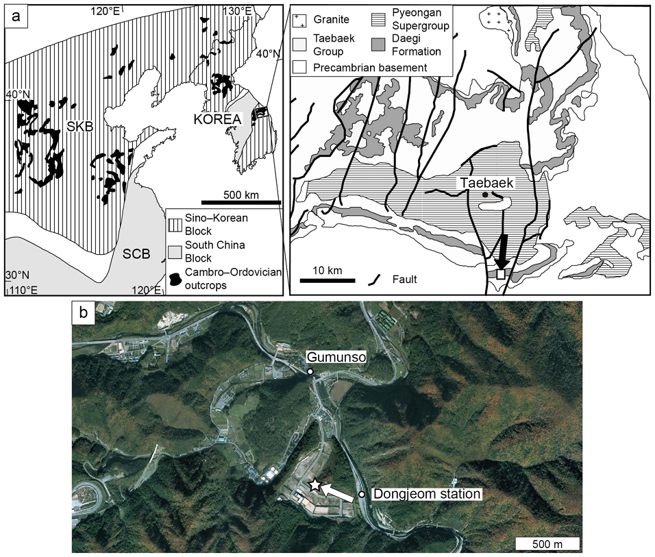
a) Tectonic elements and distribution of Cambrian-Ordovician outcrops of East Asia and simplified geologic map of the study area (arrow). Modified from Kwon et al. (2006). b) Aerial photo of the outcrop location (arrow). Satellite image is captured from kakaomap (http://map.kakao.com).
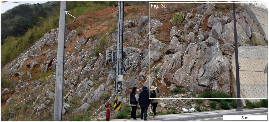
연구 지역은 최근 노출된 동점 산업단지 인근의 대기층으로서 약 25 m 층후의 석회암 노두가 비교적 연속적으로 노출되어 있다(그림 2). 생물초는 이 단면 전반에 걸쳐 발달하고 있으며, 이 연구에서는 노출된 노두 하부의 3 m 구간에 대해 조사하였다. 퇴적층의 조직적 특징을 기준으로 조사 구간 내 암상을 구분하였고, 생물초는 그 기하학적 형태 및 중규모 조직에 대해, 주위 퇴적상은 입자의 유형, 입도, 구조 및 조직에 대해 야외 정밀 관찰 및 기재하였다. 이들의 구성 요소와 미세 조직을 파악하기 위해 46개의 시료를 채취하고 다양한 면적의 연마편과 대형 박편(5.2×7.6 cm)을 제작하였다. 생물초의 경우 연마편 면을 박편화하여 총 135개의 박편을 분석하였다.
3. 대기층 미생물-석질해면류 생물초 복합체
3.1 대규모 및 중규모 구조
연구지역 대기층 노두에서는 수 십 cm~2 m 높이의 바이오험(bioherm)들이 다량으로 분포한다(그림 2, 3a). 기둥상, 구근상, 렌즈상 내지 불규칙한 외형을 가진 이들은 대체로 상부로 갈수록 너비가 커지며(그림 3a), 층리면상에서 특정 방향으로 신장된 모습은 관찰되지 않는다(그림 3b). 바이오험들은 측방으로 수 십 cm 이내의 간격을 가진 조립질 퇴적암을 사이에 두고 밀집되어 있으며, 서로 합쳐져 있는 바이오험 또한 흔하다(그림 3a-e). 생물초 내부에는 성장축을 중심으로 대칭성을 보이고 위로 볼록한 불용성 광물 심(insoluble mineral seam) 층들이 흔히 협재한다. 생물초 내부에는 수 mm 직경의 어두운 미크라이트질 덩어리가 고르게 분포하는 덩어리 조직(clotted fabric)이 관찰된다(그림 3c-d, 4a-b). 5 mm 직경 이하 크기의 아메바형 내지 신장된 형태를 보이는 어두운 미생물 덩어리는 층리에 대해 사교하는 방향 또는 수직 방향으로 서로 붙어있으며 이들의 주위에는 밝은 색의 석회 이질 퇴적물이 둘러싸고 있다(그림 4a, 4b). 국부적으로 판상 및 아치상의 미생물 크러스트가 수 cm에서 수 십 cm 높이의 렌즈상 생물초를 이룬다(그림 3a, 4c).
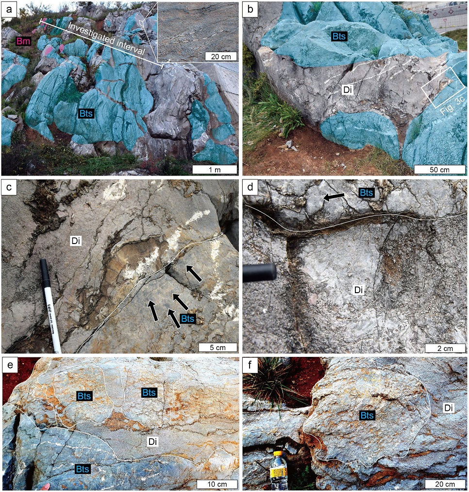
Outcrop photographs of the Daegi reef complex. a) Decimeter- to meter-scale bioherms with columnar, bulbous, lenticular to irregular geometries (blue and pink areas; Bts and Bm), forming a reef complex. Note dense development of the bioherms. An inset shows cross-stratified ooid packstone to grainstone. b) Plan view of the reef complex showing the bioherms (blue areas; Bts) separated by inter-reef deposits (Di). c) Enlargement of a rectangle in Figure 3b showing a boundary (line) between a bioherm (Bts) and an inter-reef deposit (Di). Note pervasive darker mesoclots (arrows) in the bioherms. d) Inter-reef deposit of coarse-grained limestone (Di). This rock is bounded by a bioherm (Bts) containing darker mesoclots (arrow). e-f) A few decimeter-high thrombolitic bioherms (Bts), surrounded by coarse-grained limestone (Di). Bts = thromboid-sponge bioherm, Bm = microbial crust bioherm, Di = inter-reef deposit.
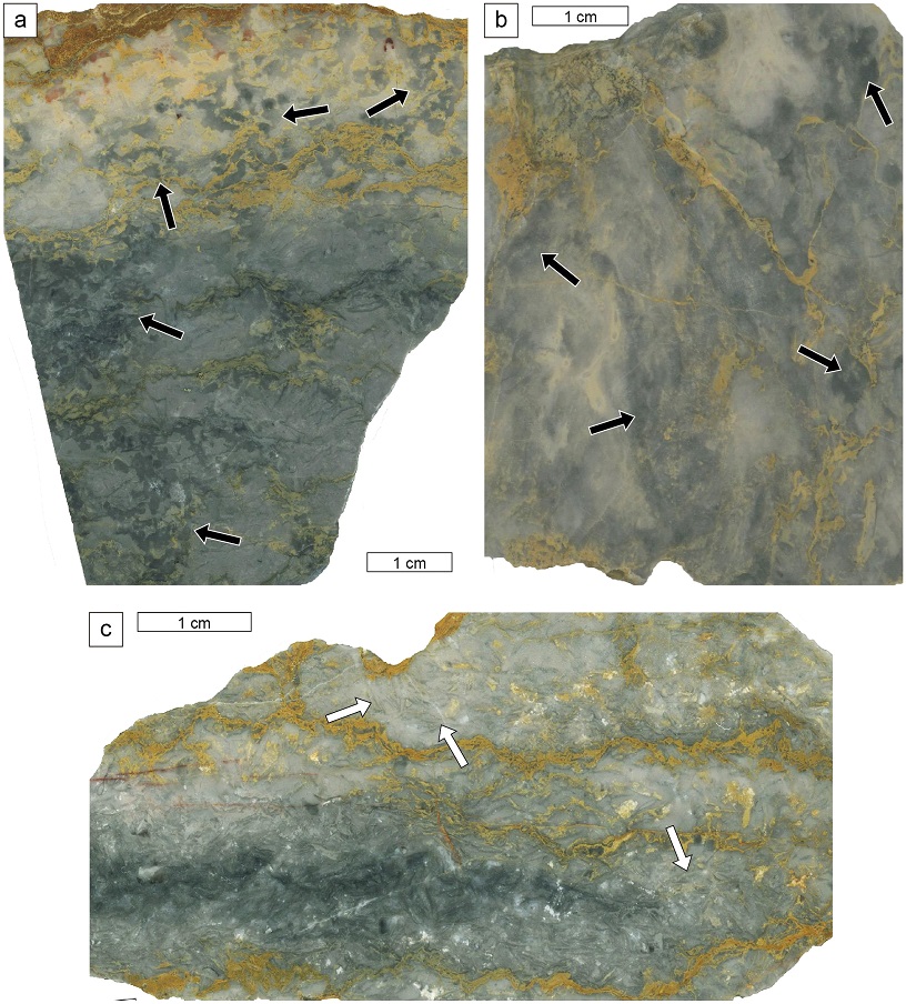
Slab photographs of the Daegi reefs. a-b) Thromboid-sponge bioherms showing millimeter-scale darker mesoclots (arrows) and the surrounding lighter micritic sediments. These mesoclots are coalesced vertically and laterally. c) Microbial crust bioherm composed predominantly of whitish, platy to arcuate crusts (arrows).
3.2 미세규모 조직: 쓰롬보이드-해면류 바운드스톤
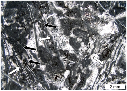
쓰롬보이드-해면류 바운드스톤은 쓰롬보이드(thromboid)가 우세하며 석질해면류 체화석, 그 파편 및 불량한 보존상태의 해면동물 골침(spicule)을 포함하는 상으로서, 구성생물 사이에는 대부분 미크라이트로 충진되어 있고 교질물은 드물다(그림 5a). 중규모 관찰 상에 어두운 미생물 덩어리로 관찰되는 쓰롬보이드는 서로 밀착되어 합쳐져 있는 500 μm에서 약 1.5 mm 크기의 마이크로스파 덩어리와 30~150 μm의 방해석 교질물로 구성된다(그림 5a). 마이크로스파 덩어리의 외형은 특정하기 어려우나 일부는 너비가 상부로 가며 넓어지는 모습이 관찰되기도 한다(그림 5a). 일부 쓰롬보이드 내에는 직경 40 μm 내외의 미크라이트질 필라멘트가 분기하거나 2차원 관찰 상에서 펠로이드가 줄지어 있는 모습처럼 보이는 석회질 미생물 Epiphyton 엽상체가 관찰되기도 한다(그림 5b, 5c).
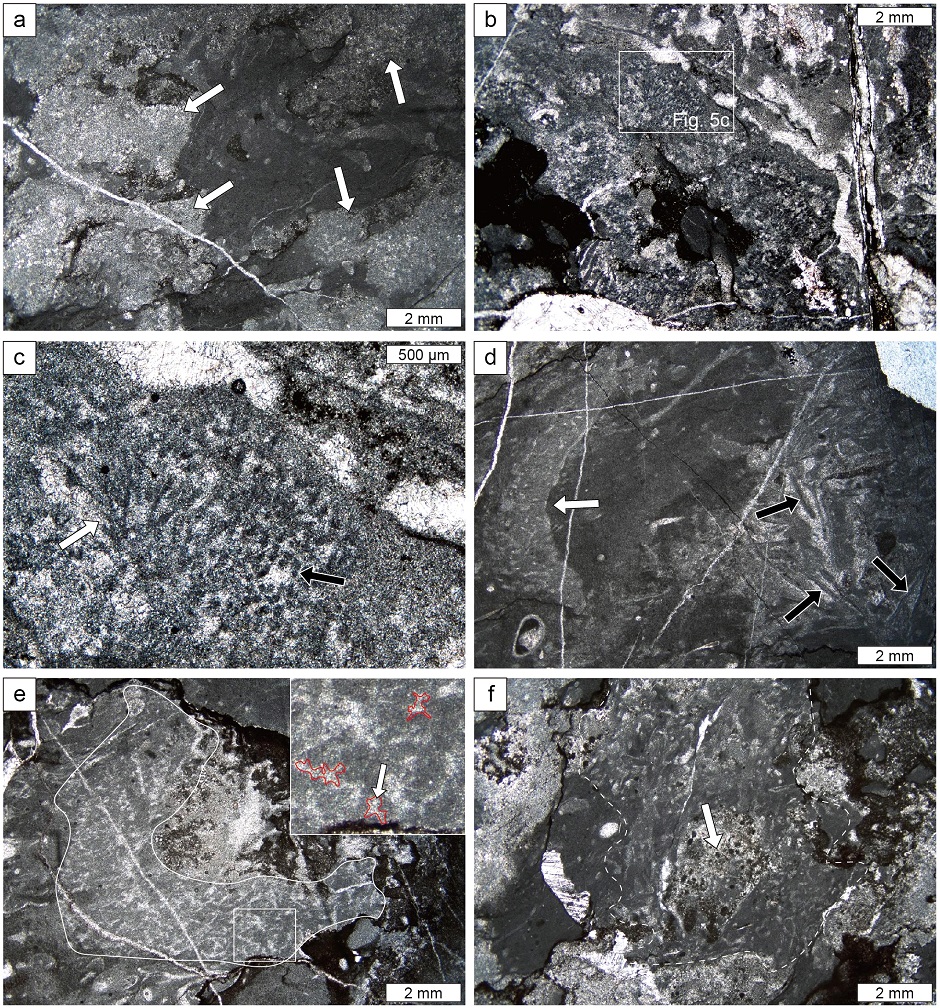
Photomicrographs of reef constituents in the thromboid-sponge boundstone. a) Millimeter-scale equidimensional thromboids (arrows), enclosed by darker micrite. b) Laterally and vertically merged Epiphyton thalli. c) Enlargement of a rectangle in Figure 5b showing bifurcation of Epiphyton filaments (white arrow). Oblique-cut filaments show arrays of peloid-like masses (black arrow). d) Oblique-cut, well-preserved lithistid sponge (white arrow) showing part of spongocoel. Obliquely to vertically aligned microbial crusts occur locally in the right (black arrows). e) Lithistid sponge skeleton (line) composed largely of straight-form spicules. Note the rectangle and its inset showing enlarged desma (red lines and arrow). f) Faintly delineated, irregular outline of a poorly preserved sponge (dashed line) with possible spongocoel (arrow). Note network-like arrangement of lighter tubules.
대기층에서 해면류는 다양한 보존 상태를 보이는데, 상대적으로 양호한 보존 상태의 화석은 중앙부에 해면위강(spongocoel)이 발달한 1~2 cm 직경의 원추형 몸체를 가지며, 곧은 골침이 망상의 배열을 이루고 미크라이트에 둘러싸인 조직을 보인다(그림 5d, 5e). 이 해면류는 다수가 층리면 방향에 대해 사교하는 방향성을 보인다(그림 5d, 5e). 해면류의 골침은 전반적으로 불규칙한 형태를 보이고 서로 연결되어 있어 개별 골침의 구분이 어려우나, 부분적으로 골침들의 접합부가 굵어지는 석질해면류의 특징인 desma 골침이 관찰되기도 한다(그림 5e). 국부적으로 보존 상태가 불량한 해면류 몸체 내에서는 펠로이드 집합처럼 보이는 구조가 관찰되기도 한다. 해면류 개체 내에서 이러한 조직들은 흔히 공존하며 서로 점이적으로 변화한다. 수 mm 크기의 보존 상태가 불량한 해면류는 망상으로 배열된 골침들의 존재를 통해 식별이 가능하나 그 외형이 아메바형 내지 불규칙하며 위강, 도관과 같은 특정 구조가 인지되지 않는다. 해면류는 대체로 단일 개체로 생물초 내에 산재해 있으며, 미크라이트에 둘러싸여 있거나 쓰롬보이드와 접해있다(그림 5d-f). 이들 외에 층리에 대해 사교하는 방향으로 비교적 일정하게 배열된 미생물 크러스트가 국부적으로 집중되어 나타난다(그림 5d).
3.3 미세규모 조직: 미생물 크러스트 바운드스톤
미생물 크러스트 바운드스톤은 100~250 μm 두께, 1~10 mm 길이를 가진 미생물 크러스트가 우세하며 풍부한 교질물이 특징적으로 나타난다(그림 4c, 6). 미생물 크러스트는 층리에 대해 수평에서 수직에 이르기까지 다양한 방향성을 보이며(그림 6), 대부분 미크라이트로 구성되나, 일정한 직경을 가진 튜브형의 석회질 미생물 Girvanella가 관찰되기도 한다. 크러스트의 윗면이나 이들의 사이 공간에는 펠로이드로 구성된 불규칙한 외형의 미생물 덩어리가 부착되어 있으며(그림 6), 미크라이트가 쌓인 경우 상하지시 구조(geopetal structure)가 관찰된다.
3.4 생물초 주위 퇴적상
대기층 생물초의 측방으로는 생쇄설물 팩스톤-그레인스톤, 일부 우이드-펠로이드 팩스톤-그레인스톤과 미생물 크러스트 팩스톤-그레인스톤이 날카로운 경계를 가지며 접한다(그림 3c-f). 이들 상은 대부분 생물초와 접하며 그 사이 공간의 형태에 따라 다양한 기하학적 형태를 보인다. 생쇄설물 팩스톤-그레인스톤은 완족류, 삼엽충, 원시해백합, 챈슬로이드, 히올리스로 주로 구성되어 있으며, 기원미상의 입자 및 펠로이드, 소량의 미생물 크러스트 또한 포함한다(그림 7a, 7b). 이들은 괴상이며 특정 퇴적구조가 관찰되지 않는다. 생파편들은 대부분 분절되어 있으며 마모되거나 부서진 형태는 드물다(그림 7a, 7b). 교질물로 충진되거나 일부 펠로이드로 충진된 생흔구조 또한 드물게 관찰된다. 입자간 공극은 다량의 방해석 교질물로 대부분 충진되어 있으나, 국부적으로 미크라이트로 채워진 공극 또한 존재한다(그림 7a, 7b). 입자간 공극의 교질물은 연변부에서는 침상, 내부에는 뭉뚝한(blocky) 형태를 보이며, 내부로 갈수록 교질물 결정의 크기가 커지는 정동 조직(drusy fabric)을 보인다(그림 7a).
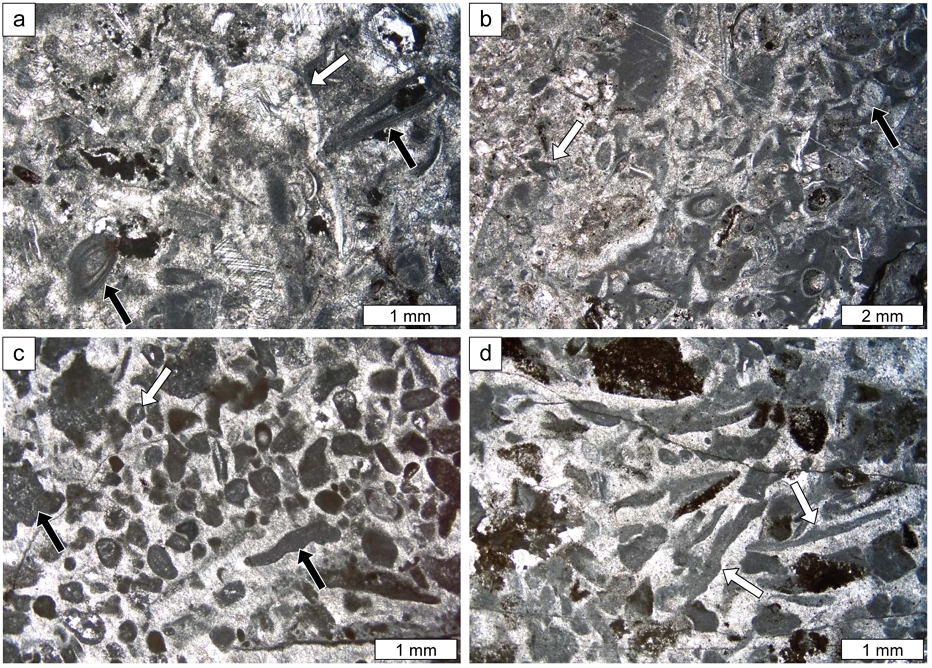
Photomicrographs of inter-reef deposits. a) Bioclastic packstone to grainstone showing grain-supported fabric with debris of trilobites (white arrow) and hyoliths (black arrows). b) Bioclastic packstone to grainstone with fragmented chancelloriids (white arrow), eocrinoids (black arrow), trilobites and hyoliths. Note higher proportion of micrite than that in Figure 7a, c) Ooid-peloid packstone to grainstone showing well-rounded and sorted micrite ooids (white arrow) and peloids with subordinate intraclasts of microbial crusts and clumps (black arrows). Small amount of micrite occurs on top of platy grains. d) Microbial crust packstone to grainstone composed largely of sub-horizontal and oblique microbial crusts (white arrows) that are sub- to well-rounded.
우이드-펠로이드 팩스톤-그레인스톤상은 분급과 원마도가 좋은 우이드와 펠로이드가 부수적인 생파편, 불규칙한 형태의 인트라클라스트, 원마도가 좋은 미생물 크러스트와 함께 입자지지 조직을 보이는 상으로서(그림 7c), 부분적으로 단일 방향성을 가진 사층리가 관찰된다(그림 3a). 우이드는 다수가 돌로마이트화 작용을 받은 우몰드(oomold)나 껍질부의 조직이 파괴된 미크라이트 우이드 또는 펠로이드로 관찰되며(그림 7c), 약 1~2 mm가량의 방사상 및 동심원상의 성장띠가 보이기도 한다. 입자간 공극은 주로 침상 및 정동 교질물로 충진되어 있으며 긴 입자의 하부 공간에 상하지시구조를 이루는 미크라이트가 소량 나타난다(그림 7c).
대체로 층리 방향에 평행하게 배열되어 있는 미생물 크러스트 팩스톤-그레인스톤상은 아원형의 원마도를 보이고 층리에 대해 무작위로 배열되어 있는 미생물 크러스트가 우세하게 나타나는 암상이며(그림 7d), 원시해백합, 삼엽충, 완족류, 히올리스, 챈슬로이드, 해면동물 골침 등의 파편이 소량으로 산출된다. 입자간 공극 충진물의 유형과 그 상대적 양은 우이드-펠로이드 팩스톤-그레인스톤과 유사하다(그림 7d).
3.5 해석
대기층 연구 구간의 미생물-우세 바이오험은 생쇄설물, 우이드-펠로이드 및 미생물 크러스트 팩스톤-그레인스톤 등 조립질 석회암과 측방으로 인접하고 있어, 수류에 의한 파괴 작용에 견딜 수 있을 정도로 견고한 골격구조가 발달한 생물초(reef) 유형으로 보인다(Wood, 1999; James and Wood, 2010). 미생물-해면류 바운드스톤에서 골격구조 사이의 공간은 생파편과 석회이질 퇴적물로 대부분 충진되어 있고 상하지시 구조와 하방으로 성장한 고착성 생물이 없어, 외부환경에서 퇴적물이 다량으로 유입될 수 있었던 개방 골격 생물초(open-frame reef) 유형이라고 판단된다(Riding, 2002). 쓰롬보이드와 Epiphyton의 골격구조 내 풍부한 양 및 상호 연결성은 이들이 생물초의 주요 건설자로 기능했으며, 단일 개체로 골격구조 내에 산재해 있는 해면류는 부차적으로 생물초 건설에 기여했을 것으로 보인다(Hong et al., 2012, 2016a). 국부적으로 나타나는 미생물 크러스트 바운드스톤은 풍부한 교질물과 상하지시 구조의 존재로 볼 때 미세골격 생물초(microframe reef)로 보이며, Girvanella가 생물초의 골격구조를 주로 형성했을 것으로 판단된다(James, 1981; Riding, 2002; Gandin et al., 2007; Hong et al., 2012, 2016a).
생물초의 측방으로 나타나는 생쇄설성 팩스톤-그레인스톤의 입자-지지 조직, 렌즈상 사층리 등 조류 기원 퇴적구조의 부재, 입자간 공극 내 구성물의 함량, 생파편의 화석생성론적 보존 상태는 이 암상이 정상파도저면(fair-weather wave base) 인근의 중에너지 내지 고에너지의 수류역학적 조건 하에서 퇴적되었음을 지시한다(Osleger and Montañez, 1996; Saltzman, 1999). 국부적으로 나타나는 우이드-펠로이드 팩스톤-그레인스톤의 풍부한 교질물의 양, 입자의 좋은 분급 및 원마도, 양호한 보존 상태의 우이드 껍질에서 나타나는 동심원상 성장 띠는 우이드와 펠로이드가 지속적으로 구를 수 있었던 고에너지 조건에서 이 상이 발달했음을 지시한다(Markello and Read, 1981; Srinivasan and Walker, 1993; Álvaro et al., 2006; Sim and Lee, 2006; Hong et al., 2016a). 미생물 크러스트 팩스톤-그레인스톤은 미생물 크러스트 생물초를 건설한 얇은 Girvanella 크러스트가 고에너지 수류에 의해 부서지며 그 인근에 쌓인 측부 퇴적물(flank deposit)로 판단된다(Rong et al., 2014; Zhang et al., 2017). 연구 구간 대기층 생물초의 밀집된 분포 양상과 측부 퇴적층의 대다수를 차지하는 생쇄설물 팩스톤-그레인스톤의 존재는 중- 내지 고에너지 조건의 교반작용(agitation)이 지속된 하부 여울 환경이 생물초를 형성한 미생물-석질해면류 군집의 발달에 적절한 서식지 조건이었음을 암시한다(cf., Woo et al., 2019).
4. 토의 및 결언
미아오링세에 발달한 미생물-고착성 동물 생물초 군집에 대한 보고는 극히 최근으로, 이들에 대한 상세한 연구는 북중국과 한반도 남부 지역을 포함하는 한중대지와 호주대륙에서만 수행되었다(Hong et al., 2012, 2016a; Kruse and Reitner, 2014; Adachi et al., 2015; Lee et al., 2016; Ezaki et al., 2017, 2020). 가장 오래된 석질해면류 체화석이 보고된 호주 북부 조지나 분지(Georgina Basin)의 란켄 석회암(Ranken Limestone)에서는 석질해면류 Rankenella와 스트로마톨라이트가 형성한 생물초가 보고되었으며 이 생물초의 측부에는 국부적으로 사층리가 발달한 생쇄설성 팩스톤-플롯스톤(floatstone)이 나타나 고에너지 환경에서 생물초가 발달했던 것으로 해석되었다(Kruse and Reitner, 2014). 란켄 석회암과 비슷한 시기에 퇴적된 중국 산동성 장샤층(Zhangxia Formation)에서는 최소 네 층준에서 석질해면류와 자포동물(cnidaria)이 소규모 쓰롬볼라이트 생물초에서 산출되며(Woo, 2009; Adachi et al., 2015; Lee et al., 2016; Ezaki et al., 2017, 2020), 이 중 상세하게 연구된 최하부의 미생물-동물 생물초는 약 150 cm 너비의 렌즈상 생물초로서, 층상의 생쇄설물-온코이드 팩스톤-그레인스톤과 생교란된 온코이드 와케스톤이 측방으로 접하는 미터 규모의 쓰롬볼라이트 내에 발달되어 있다(Lee et al., 2016). 장샤층 최하부의 생물초는 천해 조하대 환경에서 석질해면류 Rankenella와 스트로마톨라이트, 쓰롬보이드, Epiphyton 등 미생물 구성원이 생물초 골격구조를 형성했고 소량의 자포동물 Cambroctoconus가 국부적으로 생물초 발달에 기여한 것으로 보고되었다(Lee et al., 2016). 암층서 및 삼엽충 생층서 측면에서 장샤층과 매우 유사한 대기층에서는 석개재 지역에서 생물초가 보고되었는데, 대기층 하부에서 상부에 이르기까지 광역적으로 Epiphyton이 생물초 골격구조를 발달시켰으며 대부분의 생물초에서 평균 9%의 석질해면류와 보존상태가 불량한 해면류 및 국부적으로 자포동물이 생물초 건설에 참여했다고 알려져 있다(Hong et al., 2012, 2016a, 2016b). 이 생물초의 주요 발달환경은 그 상하부에서 생쇄설성 와케스톤-팩스톤이 빈번하게 분포하는 것에 비해 생쇄설성 그레인스톤은 드문 점을 근거로 저에너지의 내부 탄산염대지로 해석되었다(Hong et al., 2016a).
이 연구에서 수행된 중- 내지 고에너지 여울 환경에서 쓰롬보이드 및 Epiphyton과 석질해면류가 이룬 대기층 바이오험은 석개재 지역 대기층의 생물초와 구성 생물 측면에서 유사함에도, 주요 발달 환경 해석은 상이하다. 그러나 석개재 지역 생물초의 발달환경 해석은 노두의 불량한 보존 상태로 인하여 생물초의 공간적 밀집도에 대해 평가하지 못했으며, 그 대안으로 수행된 수직적인 현미퇴적상 분석을 통한 결과라는 맹점이 있다. 즉, 석개재 지역의 결과는 해당 지역에서 내부 탄산염대지 환경이 조성된 누적 기간이 길었음을 암시할 뿐이며, 생쇄설성 그레인스톤이 쌓인 고에너지 천해 환경이 조성된 기간은 상대적으로 짧았지만 생물초가 발달하기에 적절하지 않은 환경이었음을 의미하진 않는다. 석개재 지역에서 탄산염대지 연변부의 생쇄설성 여울 퇴적층으로 해석된 그레인스톤의 상하부에서도 대부분 쓰롬보이드-해면류 생물초가 발달되어 있는 점을 고려하면(Hong et al., 2016a), 대기층의 미생물-해면류 생물초 군집은 내부 탄산염 대지 뿐 아니라 정상파도저면 인근 및 상부의 여울을 포함한 천해 조하대 환경에서도 발달했을 것으로 추측된다(그림 8). 이 연구 결과와 유사하게 동시기 장샤층과 란켄 석회암의 생물초 역시 그 측부에 조립질 석회암상이 나타나며(Kruse and Reitner, 2014; Lee et al., 2016), 연구 구간 내 미생물-해면류 바이오험이 밀집되어 생물초 복합체를 형성한 점은 여울 환경이 이 생물초 군집의 발달에 적절한 서식지 중 하나임을 암시한다(Woo et al., 2019; 그림 8).

Environmental distribution of the Daegi reefs, which are reconstructed on the basis of Hong et al. (2016a) and this study. Note concentrated occurrences of microbial-sponge reefs in shoal environments.
북중국 장샤층과 태백층군 대기층 생물초의 구성생물은 쓰롬볼라이트, Epiphyton과 부수적인 구성생물인 석질해면류, Cambroctoconus 등 그 종류는 매우 유사하나(Hong et al., 2012, 2016a; Lee et al., 2016; Park et al., 2016; Woo et al., 2019), 두 지역 생물초에 대한 연구방법론적 차이가 뚜렷하여 동북아 지역 캄브리아기 중기 생물초의 발달 모델을 정립하는데 걸림돌로 작용해왔다. 일례로, 장샤층에서 대규모 및 중규모 중심의 분석을 통해 미생물초 복합체의 발달 양상을 제시한 Woo et al. (2019)는 쓰롬볼라이트-Epiphyton 바이오험 메가구조(megastructure)가 생쇄설성 그레인스톤이 쌓인 탄산염대지 연변부(platform margin) 환경에서 발달했으며 이러한 중 내지 고에너지 환경이 미생물초의 발달에 최적화된 환경이었다고 제안하였으나, 대기층의 생물초에 대해서는 노두의 제한 때문에 이러한 연구가 그간 불가능했다. 반대로, 장샤층의 생물초 복합체에서 해면류는 보고되지 않았는데(Woo et al., 2019), 이 화석이 육안 관찰 상에서 식별되지 않는 경우가 흔하며 대기층의 탄산염 대지 연변부 환경 생물초에서 해면류가 보고된 점을 감안하면(Hong et al., 2012, 2016a), 장샤층 생물초 복합체에서 해면류가 그 발달에 기여했을 가능성을 배제할 수 없다.
동점산업단지 인근 대기층 노두에서 생물초에 대한 다중 규모적 분석을 처음으로 수행한 본 연구는 미생물-해면류 생물초 복합체가 대기층에서 발달했음을 입증한 사례로서, 장샤층과 대기층 생물초 연구의 간극을 채울 수 있는 잠재성을 보여준다. 이 연구에서는 3 m 구간 내의 생물초에 대해 제한적으로 분석했기 때문에 현 단계에서 이들의 발달 환경을 탄산염대지 연변부의 여울인지 아니면 내부 탄산염대지의 여울환경인지 특정하기는 어려우며, 따라서 장샤층 생물초와 직접적인 비교를 하기에는 무리가 있다. 향후 대기층 노두 전반에 걸친 퇴적상 분석 및 퇴적상 조합 설정과 생물초에 대한 다중 규모 분석을 시행하면, 캄브리아기 중기 당시 동북아시아 생물초의 환경 분포 및 그 발달에 대한 조절 요인을 정교하게 이해할 수 있는 단초를 제공할 수 있으리라 판단된다. 지난 10년간 대기층과 장샤층 생물초에서 발견된 해면류의 존재가 고생대 초기 생물초의 진화사를 크게 수정한 점을 고려하면, 미생물-해면류 생물초 군집의 고생태학적 특성에 대한 향후 연구는 이에 대한 보다 고차원의 이해를 불러올 수 있을 것이라 전망한다.
Acknowledgments
이 논문은 2022년도 정부(과학기술정보통신부, 교육부)의 재원으로 한국연구재단 신진연구자지원사업(No. 2020R1C1C1007690), 대학중점연구소지원사업(No. 2019R1A6A1A03033167), 창의도전연구기반지원사업(No. 2019R1I1A1A01061336)의 지원을 받아 작성되었습니다. 유익한 지적으로 논문의 질을 향상시켜 주신 충남대 이정현 교수, 익명의 심사위원, 편집위원께 감사의 말씀을 드립니다.
References
-
Adachi, N., Kotani, A., Ezaki, Y. and Liu, J., 2015, Cambrian Series 3 lithistid sponge-microbial reefs in Shandong Province, North China: reef development after the disappearance of archaeocyaths. Lethaia, 48, 405-416.
[https://doi.org/10.1111/let.12118]

-
Álvaro, J.J., Clausen, S., Albani, A.E. and Chellai, E.H., 2006, Facies distribution of the Lower Cambrian cryptic microbial and epibenthic archaeocyathan-microbial communities, western Anti-Atlas, Morocco. Sedimentology, 53, 35-53.
[https://doi.org/10.1111/j.1365-3091.2005.00752.x]

-
Choi, D.K., 1998, The Yongwol Group (Cambrian-Ordovician) redefined: a proposal for the stratigraphic nomenclature of the Choson Supergroup. Geosciences Journal, 2, 220-234.
[https://doi.org/10.1007/BF02910166]

-
Choi, D.K., 2019, Evolution of the Taebaeksan Basin, Korea: I, early Paleozoic sedimentation in an epeiric sea and break‐up of the Sino‐Korean Craton from Gondwana. Island Arc, 28, e12275.
[https://doi.org/10.1111/iar.12275]

-
Choi, D.K., Chough, S.K., Kwon, Y.K., Lee, S.B., Woo, J., Kang, I., Lee, H.S., Lee, S.M., Son, J.W., Shinn, Y.T. and Lee, D.J., 2004, Taebaek group (Cambrian-Ordovician) in the Seokgaejae section, Taebaeksan Basin: a refined lower Paleozoic stratigraphy in Korea. Geosciences Journal, 8, 125-151.
[https://doi.org/10.1007/BF02910190]

- Chough, S.K., 2013, Geology and Sedimentology of the Korean Peninsula. Elsevier, Amsterdam, 363 p.
-
Ezaki, Y., Adachi, N., Liu, J. and Yan, Z., 2020, Cryptic growth strategies of the Cambrian coral Cambroctoconus: flexible modes of budding and growth in immediate response to available space. Palaeontology, 63, 661-674.
[https://doi.org/10.1111/pala.12481]

-
Ezaki, Y., Liu, J., Adachi, N. and Yan, Z., 2017, Microbialite development during the protracted inhibition of skeletal-dominated reefs in the Zhangxia Formation (Cambrian Series 3) in Shandong Province, North China. Palaios, 32, 559-571.
[https://doi.org/10.2110/palo.2016.097]

-
Flügel, E. and Kiessling, W., 2002, A new look at ancient reefs. In: Kiessling, W., Flügel, E., Golonka, J. (eds.), Phanerozoic Reef Patterns. SEPM No. 72, Tulsa, p. 3-10.
[https://doi.org/10.2110/pec.02.72.0003]

-
Gandin, A. and Debrenne, F., 2010, Distribution of the archaeocyath-calcimicrobial bioconstructions on the Early Cambrian shelves. Palaeoworld, 19, 222-241.
[https://doi.org/10.1016/j.palwor.2010.09.010]

-
Gandin, A., Debrenne, F. and Debrenne, M., 2007, Anatomy of the Early Cambrian ‘La Sentinella’ reef complex, Serra Scoris, SW Sardinia, Italy. In: Álvaro, J.J., Aretz, M., Boulvain, F., Munnecke, F., Vachard, D. and Vennin, E. (eds.), Palaeozoic Reefs and Bioaccumulations: Climatic and Evolutionary controls. Geological Society, 275, 29-50.
[https://doi.org/10.1144/GSL.SP.2007.275.01.03]

-
Geyer, G. and Shergold, J.H., 2000, The quest for internationally recognized divisions of Cambrian time. Episodes, 23, 188-195.
[https://doi.org/10.18814/epiiugs/2000/v23i3/006]

-
Hamdi, B., Rozanov, A.Y. and Zhuravlev, A.Y., 1995, Latest Middle Cambrian metazoan reef from northern Iran. Geological Magazine, 132, 367-373.
[https://doi.org/10.1017/S0016756800021439]

-
Hong, J., Cho, S.-H., Choh, S.-J., Woo, J. and Lee, D.-J., 2012, Middle Cambrian siliceous sponge-calcimicrobe buildups (Daegi Formation, Korea): Metazoan buildup constituents in the aftermath of the Early Cambrian extinction event. Sedimentary Geology, 253-254, 47-57.
[https://doi.org/10.1016/j.sedgeo.2012.01.011]

-
Hong, J., Lee, J.-H., Choh, S.-J. and Lee, D.-J., 2016a, Cambrian Series 3 carbonate platform of Korea dominated by microbial-sponge reefs. Sedimentary Geology, 341, 58-69.
[https://doi.org/10.1016/j.sedgeo.2016.04.012]

-
Hong, J., Choh, S.-J. and Lee, D.-J., 2016b, Distribution of chancelloriids in a middle Cambrian carbonate platform deposit, Taebaek Group, Korea. Acta Geologica Sinica, 90, 783-795.
[https://doi.org/10.1111/1755-6724.12722]

-
James, N.P., 1981, Megablocks of calcified algae in the Cow Head Breccia, western Newfoundland: Vestiges of a Cambro-Ordovician platform margin. Geological Society of America Bulletin, 92, 799-811.
[https://doi.org/10.1130/0016-7606(1981)92<799:MOCAIT>2.0.CO;2]

- James, N.P. and Wood, R., 2010, Reefs. In: James, N.P., Dalrymple, R.W. (eds.), Facies Models 4. Geological Association of Canada, p. 421-447.
-
Johns, R.A., Dattilo, B.F. and Spincer, B., 2007, Neotype and redescription of the Upper Cambrian anthaspidellid sponge, Wilbernicyathus donegani Wilson, 1950. Journal of Paleontology, 81, 435-444.
[https://doi.org/10.1666/pleo05028.1]

-
Kang, I. and Choi, D.K., 2007, Middle Cambrian trilobites and biostratigraphy of the Daegi Formation (Taebaek Group) in the Seokgaejae section, Taebaeksan Basin, Korea. Geosciences Journal, 11, 279-296.
[https://doi.org/10.1007/BF02857046]

- Kruse, P.D. and Reitner, J.R., 2014, Northern Australian microbial-metazoan reefs after the mid-Cambrian mass extinction. Memoirs of the Association of Australasian Palaeontologists, 45, 31-53.
-
Kruse, P.D. and Zhuravlev, A.Y., 2008, Middle-Late Cambrian Rankenella-Girvanella reefs of the Mila Formation, northern Iran. Canadian Journal of Earth Sciences, 45, 619-639.
[https://doi.org/10.1139/E08-016]

-
Kwon, Y.K., Chough, S.K., Choi, D.K. and Lee, D.-J., 2006, Sequence stratigraphy of the Taebaek Group (Cambrian-Ordovician), mideast Korea. Sedimentary Geology, 192, 19-55.
[https://doi.org/10.1016/j.sedgeo.2006.03.024]

-
Lee, J.-H., Hong, J., Choh, S.-J., Lee, D.-J., Woo, J. and Riding, R., 2016, Early recovery of sponge framework reefs after Cambrian archaeocyath extinction: Zhangxia Formation (early Cambrian Series 3), Shandong, North China. Palaeogeography, Palaeoclimatology, Palaeoecology, 457, 269-276.
[https://doi.org/10.1016/j.palaeo.2016.06.018]

-
Lee, J.-H. and Riding, R., 2018, Marine oxygenation, lithistid sponges, and the early history of Paleozoic skeletal reefs. Earth-Science Reviews, 181, 98-121.
[https://doi.org/10.1016/j.earscirev.2018.04.003]

-
Markello, J.R. and Read, J.F., 1981, Carbonate ramp‐to-deeper shale shelf transitions of an Upper Cambrian intrashelf basin, Nolichucky Formation, Southwest Virginia Appalachians. Sedimentology, 28, 573-597.
[https://doi.org/10.1111/j.1365-3091.1981.tb01702.x]

-
McKenzie, N.R., Hughes, N.C., Myrow, P.M., Choi, D.K. and Park, T.-Y., 2011, Trilobites and zircons link north China with the eastern Himalaya during the Cambrian. Geology, 39, 591-594.
[https://doi.org/10.1130/G31838.1]

-
Oslegar, D.A. and Montañez, I.P., 1996, Cross-platform architecture of a sequence boundary in mixed siliciclastic-carbonate lithofacies, Middle Cambrian, southern Great Basin, USA. Sedimentology, 43, 197-217.
[https://doi.org/10.1046/j.1365-3091.1996.d01-13.x]

-
Park, T.Y., Han, Z., Bai, Z. and Choi, D.K., 2008, Two middle Cambrian trilobite genera, Cyclolorenzella Kobayashi, 1960 and Jiulongshania gen. nov., from Korea and China. Alcheringa, 32, 247-269.
[https://doi.org/10.1080/03115510802096135]

-
Park, T.-Y.S., Kihm, J.-H., Woo, J., Zhen, Y.-Y., Engelbretsen, M., Hong, J., Choh, S.-J. and Lee, D.-J., 2016, Cambrian stem-group cnidarians with a new species from the Cambrian Series 3 of the Taebaeksan Basin. Acta Geologica Sinica, 90, 827-837.
[https://doi.org/10.1111/1755-6724.12726]

-
Riding, R., 2002, Structure and composition of organic reefs and carbonate mud mounds: concepts and categories. Earth-Science Reviews, 58, 163-231.
[https://doi.org/10.1016/S0012-8252(01)00089-7]

-
Rong, H., Jiao, Y., Wang, Y., Wu, L. and Wang, R., 2014, Distribution and geologic significance of Girvanella within the Yijiangfang Ordovician reef complexes in the Bachu area, West Tarim Basin, China. Facies, 60, 685-702.
[https://doi.org/10.1007/s10347-013-0394-9]

-
Rowland, S.M. and Shapiro, R.S., 2002, Reef patterns and environmental influences in the Cambrian and earliest Ordovician. In: Kiessling, W., Flügel, E., Golonka, J. (eds.), Phanerozoic Reef Patterns. SEPM No. 72, Tulsa, p. 95-128.
[https://doi.org/10.2110/pec.02.72.0095]

-
Saltzman, M.R., 1999, Upper Cambrian carbonate platform evolution, Elvinia and Taenicephalus zones (Pterocephaliid-Ptychaspid Biomere boundary), northwestern Wyoming. Journal of Sedimentary Research, 69, 926-938.
[https://doi.org/10.2110/jsr.69.926]

-
Servais, T., Owen, A.W., Harper, D.A.T., Kröger, B. and Munnecke, A., 2010, The Great Ordovician Biodiversification Event (GOBE): the palaeoecological dimension. Palaeogeography, Palaeoclimatology, Palaeoecology, 294, 99-119.
[https://doi.org/10.1016/j.palaeo.2010.05.031]

-
Shapiro, R.S. and Rigby, J.K., 2004, First occurrence of an in situ anthaspidellid sponge in a dendrolite mound (Upper Cambrian; Great Basin, USA). Journal of Paleontology, 78, 645-650.
[https://doi.org/10.1666/0022-3360(2004)078<0645:FOOAIS>2.0.CO;2]

-
Sim, M.S. and Lee, Y.I., 2006, Sequence stratigraphy of the Middle Cambrian Daegi Formation (Korea), and its bearing on the regional stratigraphic correlation. Sedimentary Geology, 191, 151-169.
[https://doi.org/10.1016/j.sedgeo.2006.03.016]

-
Srinivasan, K. and Walker, K.R., 1993, Sequence stratigraphy of an intrashelf basin carbonate ramp to rimmed platform transition: Maryville Limestone (Middle Cambrian), southern Appalachians. Geological Society of America Bulletin, 105, 883-896.
[https://doi.org/10.1130/0016-7606(1993)105<0883:SSOAIB>2.3.CO;2]

-
Webby, B.D., 2002, Patterns of Ordovician reef development. In: Kiessling, W., Flügel, E., Golonka, J. (eds.), Phanerozoic Reef Patterns. SEPM No. 72, Tulsa, p. 129-180.
[https://doi.org/10.2110/pec.02.72.0129]

- Woo, J., 2009, Depositional environments and microbial sedimentation of the Zhangxia Formation (Middle Cambrian). Ph.D. thesis, Seoul National University, Seoul, 231 p.
-
Woo, J., Kim, Y.-H.S. and Chough, S.K., 2019, Facies and platform development of a microbe-dominated carbonate platform The Zhangxia Formation (Drumian, Cambrian Series 3), Shandong Province, China. Geological Journal, 54, 1993-2015.
[https://doi.org/10.1002/gj.3274]

- Wood, R., 1999, Reef Evolution. Oxford University Press, Oxford, 414 p.
-
Zhang, M., Hong, J., Choh, S.-J. and Lee, D.-J., 2017, Thrombolite reefs with archaeocyaths from the Xiannüdong Formation (Cambrian Series 2), Sichuan, China: implications for early Paleozoic bioconstruction. Geosciences Journal, 21, 655-666.
[https://doi.org/10.1007/s12303-017-0011-y]


